Clear epidermal cells of an onion, allium cepa, in a single layer. Scanning electron micrograph (sem) of adipocytes (ad) membrane structure and function prokaryotic cells:

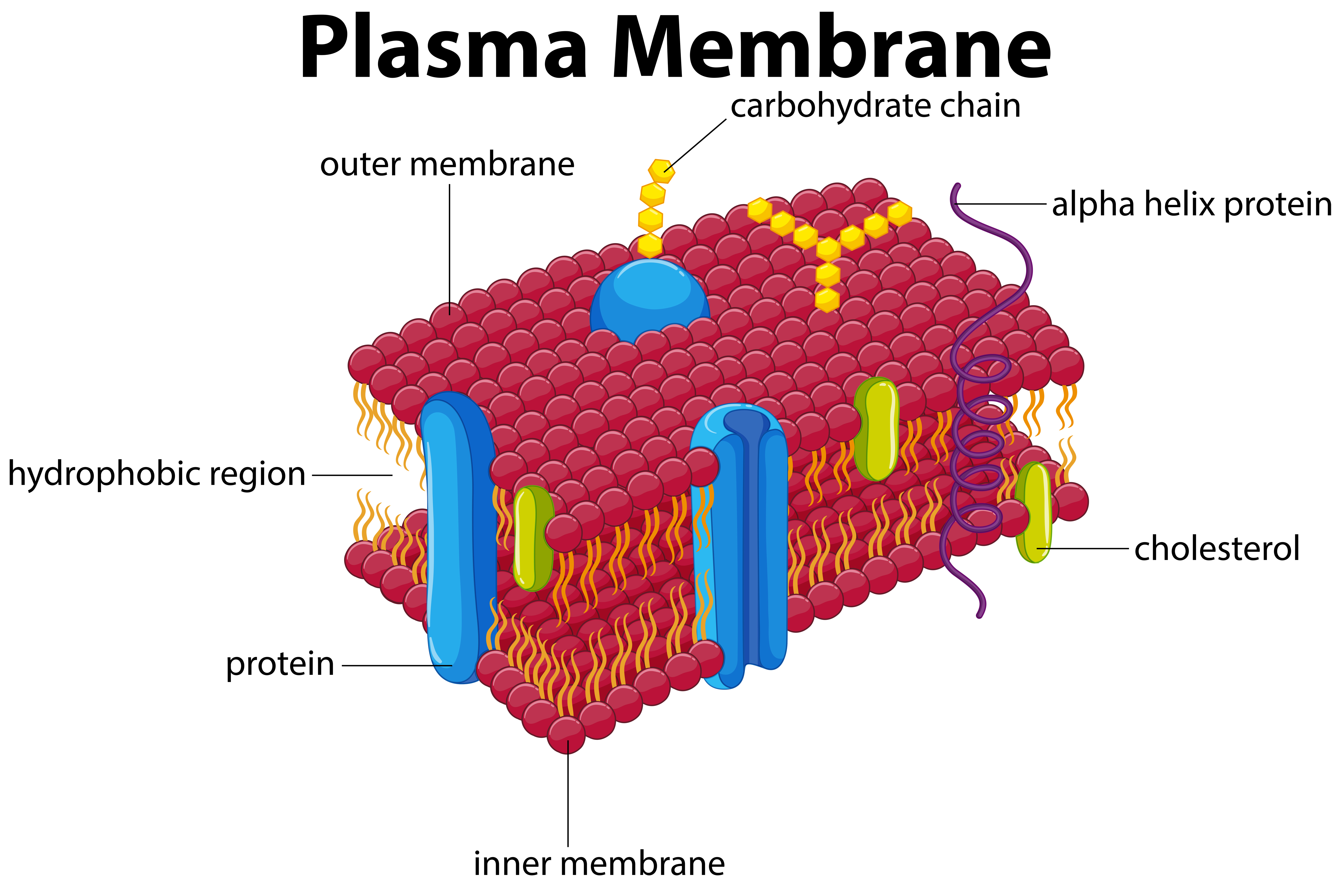
The cell membrane is an extremely pliable structure composed primarily of two layers of phospholipids (a “bilayer”).
Cell membrane diagram labeled. Scanning electron micrograph (sem) of adipocytes (ad) membrane structure and function prokaryotic cells: It protects the integrity of the cell along with supporting the cell and helping to maintain the cell�s shape. The cell membrane regulates the transport of materials entering and exiting the cell.
Clear epidermal cells of an onion, allium cepa, in a single layer. The cell membrane consists of a lipid bilayer that is semipermeable. There are no organelles in the prokaryotic cells, i.e., they have no internal membrane systems.
The lipid tails of one layer face the lipid tails of the other layer, meeting at the interface of the two layers. Help students visualize the structure of a cell membrane by using an analogy. Hence the bacterial cell structure lacks a nucleus.
Use for everything except reselling item itself. The cell membrane consists of two adjacent layers of phospholipids. Terms in this set (6) channel protein.
In this diagram of a cell membrane, the object labeled (b) is a m2 o carbo carbohydrate side chain. Learn about the structures of cell membrane or plasma membrane with this collection of printable cell membrane diagrams. The exact mix or ratio of proteins and lipids can vary depending on the function of a.
Proteins and lipids are the major components of the cell membrane. Label a cell labelled diagram. Cell and membrane transport labeled.
In a normal cell, membrane transport is vital for the movement of glucose and amino acids into the cells for the production of energy and protein synthesis,, respectively. This layer of cellulose fiber gives the cell most of its support and structure. Label the cell membrane labelled diagram.
Cell membrane the cell membrane is the outer coating of the cell and contains the cytoplasm, substances within it and the organelle. Structure and composition of the cell membrane. Dec 13, 2021 · simple cuboidal epithelium:
The structure labeled b in the diagram is an example of a n. Diagram of the human cell illustrating the different parts of the cell. While lipids help to give membranes their.
Students learn by coloring and identifying each component that. The correctly labelled diagram is (a) nucleus. Cell membrane with labeled educational structure scheme vector illustration.
Since the cell membrane is made up of a lipid bilayer with proteins attached on. Cell membrane (plasma membrane) =. Anatomical closeup drawing with cross section element.
The formation of cell membranes is crucial to life. Cholesterol and various proteins are also embedded within the. Controls passage of materials in and out of the cell chromosomes condense, nuclear membrane dissolves.mitosis:
The cell membrane is an extremely pliable structure composed primarily of two layers of phospholipids (a “bilayer”). The cell membrane is a multifaceted membrane that envelopes a cell�s cytoplasm. Hole or tunnel that particles may pass through to go in / out of cell.
A labeled diagram of the animal cell and its organelles biology wise. Cell wall a thick, rigid membrane that surrounds a plant cell. This cell membrane provides a protective barrier around the cell and regulates which materials can pass in or out.
For the human cells, the nuclear materials are separated from the cytoplasm by a membrane known as the nuclear membrane. Facilitated diffusion through cell membrane (with diagram) a variety of compounds including sugars and amino acids pass through the plasma membrane and into the cell at a much higher rate than would be expected on the basis of their size, charge, distribution coefficient, or magnitude of the concentration gradient. K g1 g2 g3 g4 g5 g6 g7 g8 g9 g10 g11 g12 university animal.
To study the structure of the onion epidermal cell, with particular. Label the cell membrane labelled diagram. Labeled diagram simple cuboidal epithelial cells are shaped like cubes, and the nucleus of each cell is large and located close to the center of the cell.
Known as retinal (a pigment also found in the human eye) act similar to chlorophyll. Labeled diagram of plasma membrane luxury in the cell membrane plasma membrane phospholipid bila plasma membrane cell membrane cell membrane coloring worksheet. Channel protein, cholesterol, external cell environment, hydrophilic (water loving) part of phospholipid bilayer, peripheral protein, internal environment of the cell, hydrophobic (water fearing) part of phospholipid bilayer, glycolipid.
Start studying labeling a cell membrane. (3 marks) d draw and label a few onion epidermal cells observed under the microscope. Because the bacteria cell structure lacks a nuclear membrane, they are group as prokaryotic cells different from the cells with nuclear membranes known as eukaryotic cells.
Carbohydrate, globular protein or cholesterol location visualization. Practice labeling the parts of the cell membrane. Both humans and onions are eukaryotes, organisms with relatively large, complicated.
The cell membrane, also called the plasma membrane, is found in all cells and separates the interior of the cell from the outside environment. The phospholipid heads face outward, one layer exposed to the interior of the cell and one layer exposed to the exterior (figure 2). Membrane structure and function all cells have a plasma or cell membrane , which contains the cell.
Drawing diagram of a cell membrane : If all the cells in an organism suddenly die the organism itself dies as well. Learn vocabulary, terms, and more with flashcards, games, and other study tools.